World of Urology
World of Urology

World of Urology is a group of the Best Urologists in Bangalore who are all leading specialists in their fields. Together, we provide comprehensive services of the highest standard for prostate cancer, bladder, and kidney problems.
Transforming lives by providing 360° diagnosis & surgical care
World of Urology is a Certified, well-trained and committed group of 10 Best Urologist in Bangalore providing 360 degree URO surgical and kidney transplant care.
Expertise in Robotic Uro Oncology, Robotic Urology, 3D Laparoscopy, Laser Urology, Kidney transplant surgery, Penile Implants, Kidney operation, kidney cancer treatments, Uterus removal surgery, Robotic surgery for Prostate cancer, Laser enabled kidney stone & Prostate surgery, Complex Urethral reconstruction including redo hypospadias/Exstrophy Epispadias single stage repair, Pediatric Urology, Minimal Invasive Surgery for Urinary Incontinence and URO gynecology problems.
Services
Robotic Pyeloplasty
Robot-Assisted Laparoscopic Pyeloplasty
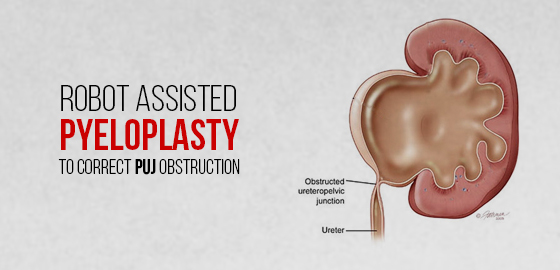
WORLD OF UROLOGY – WHAT IS A PYELOPLASTY?
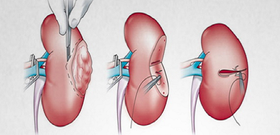
A Pyeloplasty is an operation done to correct PUJ obstruction, wherein the tube that connects the kidney to the bladder (ureter) is narrowed at its junction with the kidney and does? allow the kidney to drain properly and hence, affects its function.
HOW IT IS DONE?
The operation can be done either by conventional Open method or by Keyhole surgery, with or without the help of a Robot.
WHAT IS AN OPEN PYELOPLASTY?
Open Pyeloplasty is whereby a surgeon uses an incision of approximately 12-18cm in length to carry out reconstruction of your kidneys drainage system to improve kidney function.
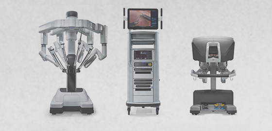
WHAT IS ROBOTIC ASSISTED SURGERY?
This is a technique whereby a robotic console is placed beside you. Attached to the console are 3 robotic arms; two for instrument attachments and the other arm for a high magnification 3D camera to allow the surgeon to see within your abdomen (tummy). The two robotic arms have the ability to hold various instruments attached to them to allow the surgeon to carry out your operation. The instruments are approximately 7mm in width.
The instruments have a greater range of movement than a human hand does; and because of their size and the ability to view the operation in 3 dimensions; this allows the surgeon to carry out surgery in a small space.
Previous surgeons made larger incisions to be able to perform most operations. With robotic surgery the instruments are placed onto the robotic arms through small port holes into your abdomen; the surgeon sits in the same room but away from the patient and along with a bedside surgeon next to the patient, is able to carry out more controlled and precise movements using robotic assistance.
Advantages :
- Avoids open surgery and the resulting big scar
- Considerably less bleeding
- Precise surgery resulting in better outcomes
- Less pain after the operation.
- Shorter hospital stays.
- Quicker full recovery and earlier return to work.
WHAT ARE THE RISKS OF THE PROCEDURE?
As in any surgery, there are a few risks of which the common ones are:
- During port placement: bleeding, and damage to structures inside the abdomen (tummy)-this is minimized by placing ports under vision.
- During the operation: bleeding, conversion to open surgery, injury to structures in the abdomen
- During exit from abdomen: bleeding
- After the operation: bleeding, infection, hernia at port site, blood clots in legs which can migrate, shoulder tip pain
- Leakage of urine at the kidney joining the ureter generally settles by itself.
DOES THE ROBOT DO THE SURGERY?
No, the surgeon does the operation. The robot is an instrument that allows the surgeon to operate in small spaces in the body. It essentially makes the surgeon’s hands two seven-millimetre instruments. The robot is controlled by the surgeon and does not work on its own.
HOW MUCH PAIN WILL I BE IN?
Since the surgery is done through a small incision, most patients experience much less post-procedure pain than with open surgery. Patients tend to need much less pain medication. After one week, most are feeling no pain at all. Also, there is a decreased risk of post-operative hernias.
WHEN CAN I EXERCISE?
Light walking is encouraged right after the procedure. After 2 weeks, jogging and aerobic exercise is permitted. After four weeks, heavy lifting can resume.
CAN I SHOWER OR BATH?
Yes, the stitches in your tummy are dissolvable; we just asked that you rinse thoroughly the soap from your body as this may irritate the wounds and that you pat yourself completely dry.
WHEN CAN I DRIVE?
When you are comfortable to do so and when able to make an emergency stop.
World of Urology – RAVH

Daycare RAVH (Robotic Assisted Vaginal Hysterectomy)
Hysterectomy is the surgical removal of the uterus. Hysterectomy can be performed for a variety of reasons and may also involve the removal of other organs and tissues, including ovaries and/or fallopian tubes. This surgery traditionally done via open surgical method involved a large incision in the abdomen and postoperative significant pain. With the advent of minimal access surgery, the surgery can be performed safely with minimal blood loss and early recovery. Robotics has added a new dimension to minimal access surgery by giving the surgeon a 3D
perception and magnification of the organs that help in precision surgery to add to the safety of the patient.
WHY IS A HYSTERECTOMY PERFORMED?
Hysterectomies may be performed to treat conditions such as:
- Uterine fibroids (Leiomyomas) which are causing intense pain & bleeding.
- Severe Endometriosis which is characterized by the growth of uterine tissue outside the uterus.
- Cancer or pre-cancer of the uterus, cervical or uterine cancer.
- Uterine prolapse.
- Abnormal vaginal bleeding is not being controlled by other treatment methods.
- Increased intensity of pelvic pain which is not being controlled by other treatment methods.
THE SURGICAL APPROACH
Hysterectomy is one of the most common surgeries performed by gynaecologists and uro gynaecologists worldwide. Increasingly more and more surgeons across the world have started using the Da-Vinci robot for gynaecological surgeries.
The magnified, 3-dimensional view allows for incredible precision, flexibility and control. The surgeon passes the instruments through small abdominal incisions. The surgeon can control all these movements from a console that, in turn, provides a greater level of comfort for him/her. In addition, the articulating arms offer 7 degrees of movement for precise movements enabling the surgeon to make accurate &safe cuts. Last but not least, this method also drastically decreases the chances of the surgeon experiencing tiredness or fatigue at the end of the day thereby helping reduce the possibility of human error as well.
Patients undergoing a minimally invasive hysterectomy are also likely to experience lower levels of pain, experience decreased blood loss, lesser chances of contracting infections, less scarring as well as quicker recovery time. Patients can recover very quickly from all sorts of difficult/complex surgeries while being able to resume normal daily activities more quickly than one could after undergoing open surgery. All this is possible while also maintaining relatively good outcomes and low complication rates.
A robotic hysterectomy may be recommended even to severely obese patients where an open or laparoscopic hysterectomy would be difficult to perform.
BENEFITS TO THE PATIENT:-
- Lower levels of pain.
- Lesser chances of contracting infections.
- Decreased blood loss.
- Less scarring.
- Shorter hospital stays.
- Quicker recovery time to resume normal daily activities.
All this is possible while also maintaining relatively good outcomes and low complication rates.
THE PROCEDURE
A robotic-assisted vaginal hysterectomy is performed under General anaesthesia. More often than not, the average operating time ranges between 1-3 hours. That said, operative time tends to differ from individual to individual.
In some cases, a urinary catheter might be inserted into the patient to empty his/her bladder. Thereon, approximately 3 to 5 small keyhole (< 1cm) incisions are made in the abdomen and slender surgical instruments are inserted through them. The instruments are then docked onto the robot. The surgeon remotely controls the robot with the aid of a console located a short distance away from the patient where the magnified operating field can be viewed in 3-D. With the help of this console, the surgeon will be able to control the surgical instruments in order to remove the uterus.
Depending on the patient’s condition, the surgeon might also proceed to remove one or both ovaries as well as the fallopian tubes.
World of Urology – Nephron Sparing Surgery
Renal tumours are most commonly detected during health checkups or when one presents for some other ailment and investigations are performed. Hence the importance of periodic health check-ups cannot be stressed enough.
Commonly mentioned symptoms of kidney tumours including blood in urine, flank pain and a palpable mass are present only in advanced cases where a complete cure is not feasible.
Earlier the diagnosis of kidney tumours involved surgical removal of the entire kidney. In recent times the need to remove the entire kidney has been negated. Nephron-sparing surgery has ushered in a sea change in the management of this otherwise fatal disease. It involves removing the tumour with a rim of normal tissue thus getting rid of the cancerous tissue and sparing an others normal kidney.
Again the advent of minimally invasive surgical procedures like laparoscopy and robotic surgery has substantially reduced the size of the incision, the recovery period and the time to return to normal life.
NEPHRON-SPARING SURGERY (PARTIAL NEPHRECTOMY)
There are approximately 1 million nephrons in each human kidney and together they are responsible for various functions of the kidney including maintaining fluid balance in the body, filtering waste material, producing haemoglobin, promoting bone health, regulating blood pressure and much more.
When a tumour is detected in a kidney, the ability to remove just that portion of the kidney allows a significant percentage of normal kidney function to be retained, which is even more important in a diabetic or a hypertensive who might in due course of time undergo further deterioration in kidney function due to these diseases. Even in a normal individual preserving kidney function by performing such surgery has produced wonderful results.
SURGICAL PROCEDURE
This involves careful dissection of the blood vessels (renal artery and vein) and their branches and then selectively clamping that branch of the vessels which feeds the area of the tumour. Then an incision is made around the tumour excising the tumour with a small rim of normal renal tissue. This part is then sent to the pathology department – a process known as a frozen section – where a quick examination reveals whether the tumour is malignant and if yes, has the tumour has been completely excised. This allows complete and total resection of the tumour allowing a radical cure.
ROBOTIC PARTIAL NEPHRECTOMY
Open surgical procedures of Partial Nephrectomy resulted in large incisions with significantly delayed recovery and post-operative pain.
The Da Vinci Robotic system allows a minimally invasive approach which under general anaesthesia allows 3-4 robotic arms to be inserted through 1 cm incisions and allows the dexterity and manoeuvrability of the human hand to be replicated inside the body with minimal incisions. Visualising the entire field with 3D vision allows for wonderful precision, reduced blood loss and absolute surgical comfort and ease.
The post-operative pain is reduced by a great degree and so is the recovery period which enables a very early return to normal life.
This procedure is also a boon for the operating surgeon as it’s performed on a console which allows him greater comfort and ease thus reducing surgical time, room for error and surgical fatigue.
Address: No 156, Orchard Grande, 4 th Cross, 8th B-Main Sadashivanagar, Bangalore 560080
Location:- https://goo.gl/maps/oVgbWK1gDSkEkm5i6
visit are blog: https://www.worldofurology.in/blog
resources URL: https://www.worldofurology.in/
Twitter: https://twitter.com/worldofurology
Facebook: https://www.facebook.com/WorldUro/
Instagram:- https://www.instagram.com/worlduro/
Linkedin:- https://www.linkedin.com/company/world-of-urology/


Comments
Post a Comment